PhD thesis, Katholieke Universiteit Leuven and Eindhoven University of Technology (TU/e), 15 February 2010, 171 pages.
The volume of image guided interventions and therapy is rapidly increasing, because of associated better clinical outcome and reduced patient strain. During such minimally invasive medical treatment the clinician relies on radiological images, often produced in real-time, to guide the intervention. Prior to treatment, diagnosis and intervention planning is frequently performed using tomographical images, which provide a detailed representation of the patient's anatomy and pathology. In this thesis the fusion of those different types of images is presented, in order to provide the clinician with more relevant data to guide the procedure. Since the fusion is performed during the clinical intervention, it is essential that the technical steps can be executed within limited time. Furthermore, it is vital that the resulting fused representations are easy to interpret. The technical approaches that are described here to achieve this goal comprise fast and intuitive visualization of the fused data and rapid co-registration of multiple image datasets.
In order to achieve an optimal performance the parallel computation power of the graphics processing unit (GPU) has been exploited in the visualization and registration algorithms. Regarding visualization, a dedicated direct volume rendering approach was developed, taking the particularities of the GPU into account. This volume rendering technique has been applied in the efficient fused visualization of multiple datasets, and in the interactive rendering for autostereoscopic displays. An elastic B-spline driven registration method has been mapped on the GPU to accomplish minimal computation times. Furthermore, registration algorithms especially designed for peri-interventional usage were examined. A registration method only using the geometry information of the X-ray C-arm system has been described, and a dedicated registration algorithm targeted at real-time vascular images has been developed.
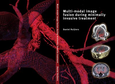
The proposed techniques have been validated individually, and have been evaluated together in three concrete clinical applications: multi-modal roadmapping for neuro-vascular treatment, multi-modal needle puncture planning and tracking, and CT fusion with X-ray angiography for stent placement within coronary artery disease treatment.
Academic supervisors:
